This article based on Knowles Precision Devices blog provides an overview of trends in medical electronics and related use of passive electronic components.
Digital and connected healthcare methods are getting better and better at harnessing the full potential of today’s advanced medical technologies and popular consumer devices.
This combination is evolving standard healthcare delivery for the digital age. While these technologies still pose privacy, security and access challenges, they’ve made significant strides. The vision of a less hospital-centric, and more patient-centric, system is starting to crystallize.
We’ll also scratch the surface on three trends that are improving medical imaging technologies, and by extension, diagnostics, patient outcomes and access to care. We’ll use magnetic resonance imaging (MRI), which leverages radio frequency (RF) signals, to contextualize these trends.
Medical Electronics Technology Trends
Here are a few areas where outcomes are improving as technology moves closer to patients.
Wearables
In a healthcare context, wearable technology includes devices that patients or consumers can wear to collect personal data on certain health and exercise metrics. They typically feature sensing technologies that detect information and transmit back to smartphone applications where data can be viewed, analyzed and managed. Growing consumer interest in wearable fitness trackers paired with more ubiquitous network coverage (e.g., smartphones) is setting the scene for more personalized and timely healthcare guidance in terms of monitoring, diagnostics and therapeutics.
Hearables
As a subcategory, hearables encompass a wide variety of wearable technology used for listening and/or monitoring (e.g., health, activity). In the healthcare space, many of these devices are positioned as wellness technologies, but they serve as a foundation for future FDA-approved medical devices. Biofeedback sensors placed in the ear could offer more information on key health parameters. Hearables are expanding the options for those who need hearing aids, but they’re also another source of information like heart rate, blood oxygen saturation and blood pressure.
Remote Patient Monitoring
As remote patient monitoring (RPM) devices evolve into a more effective and more affordable option, they’re becoming more prevalent in patient care plans. RPM devices electronically transmit data between patients and healthcare professionals, on a continuous or periodic basis, even when the patient is outside of a traditional clinical setting. With more information, healthcare professionals have a more accurate picture of a patient’s health and may require less in-person or virtual visits—extending results-driven care to different contexts with less adverse effects or negative outcomes.
Point of Care Diagnosis
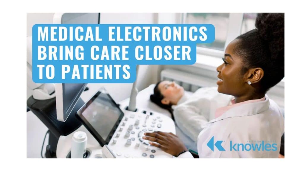
After decades of technological development, point-of-care ultrasound (POCUS) is a widely accessible and affordable method of performing advanced diagnostic services. Patient assessment used to rely on technology that required its own suite for proper function, like computed tomography (CT) scans or magnetic resonance imaging (MRI). These imaging modalities are sensitive enough for diagnosis, but they can be time-consuming and physically demanding for critically ill patients. With the invention of ultrasound technology and the availability of better transducers, clearer images, and real-time display, diagnostics can occur at a patient’s bedside regardless of the setting. Today, healthcare professionals can more swiftly identify patient conditions and make more informed decisions about their care. This improves outcomes, avoids invasive procedures and, ultimately, decreases costs.
Smart Implants
Smart implants, including pacemakers and neuromodulators, are designed to treat diseases, track patient information and/or stimulate regular system functions. With the right combination of sensors and circuits, these devices go beyond replacing organs or tissues. They add functions that weren’t originally present or restore functions that were lost. For smart implants to flourish, designs need be simple, highly sensitive, cost effective and have favorable clinical effects (i.e., avoid harm and adverse effects).
Medical Imaging Overview
Medical professionals rely on a variety of medical imaging techniques to non-invasively visualize internal body structures and functions. These techniques are useful for diagnosing diseases and injuries, monitoring treatment performance and surgical planning. Each imaging modality is designed with different clinical use cases in mind (Figure 1).
| Technology | Signal* | Frequency | Applications |
| Ultrasound | Sound | 10’s of KHz+ | Blood vessels, muscles, tendons and blood flow |
| MRI | RF Signals | 10’s of MHz+ | Soft tissue |
| Endoscopy | Visible and IR | 100’s of GHz+ | Gastroscopy, brochoscopy, uretroscopy, neuroscopy |
| OCT | Opthalmology, dermatology, cardiology, oncology | ||
| X-Ray | X-Ray Radiation | 10’s of PHz+ | Bones, Soft Tissue |
| CT-Scan | Head, neck, lungs, cardiac, abdominal, bones, ligaments | ||
| PET | Gamma Radiation | 10’s of EHz+ | Oncology |
| SPEC | Oncology |
* Signals range from sound waves at 20kHz to Gamma Radiation at Exa-Hertz
Combining Imaging Modalities
Hybrid imaging technologies leverage the strength of a combination of techniques to produce highly detailed views of the body. Medical professionals use these images to diagnose and treat patients more effectively.
For example, PET/MRI scans integrate positron emission tomography (PET) scans and MRI scans. MRI offers detailed images of internal bodily structures and their functions while PET highlights abnormalities using tracers. This combination is particularly helpful in care related to Alzheimer’s disease, epilepsy and brain tumors. Historically, it wasn’t possible to integrate PET and MRI because MRI’s strong magnets interfered with PET’s imaging detectors. Scans were taken separately and merged, which involved complex image processing and potential data loss. According to Stanford Medicine, the PET/MRI combination is more accurate, safer and more convenient than performing separate scans.
Increasing Imaging System Performance
Performance improvements lead to better image quality and more accurate information for diagnostic and treatment purposes. For example, researchers have access to MRI systems with field strength up to 7T now. This performance improvement enhances signal-to-noise ratio (SNR), so imaging results are clearer and more detailed. There’s also a push to make MRI receivers more digital in nature. With higher resolution and higher frequency analog-to-digital converters (ADCs) available, there’s an opportunity to shift the ADC to the RF coil, which can reduce noise and increase SNR when power consumption is managed appropriately. Similar benefits can also be achieved by adding more individual RF coils to the system. Focusing on performance translates to improving elements of patient experience like scan times and cost too.
Designing Imaging Equipment for Portability
By design, some patient assessment and treatment equipment started out in controlled environments for proper function (e.g., MRI suite). Computed tomography (CT) and magnetic resonance imaging (MRI) are great examples. While these imaging techniques are effective for diagnosis, they can be physically demanding for critically ill patients. Technological development is moving these diagnostic services to where patients are.
For traditionally stationary devices like MRI machines, designing for portability requires consideration for factors like size and weight, power, magnetic field strength, cost, image quality and safety. At the components level, selections like high-performance capacitors are important for stable and efficient power generation and signal processing in a smaller, portable footprint.